A patient is told from the outset that his vision has a ceiling. Not because of the prescription, which can be corrected, but because his eye learned to see poorly in childhood and that does not undo. His estimated potential is 20/40, which in practice means half of what a person with normal vision sees.
The day after surgery he was seeing 20/30 in one eye and 20/25 in the other, with nothing in front of them. Better without glasses than he had ever seen with them.
This article explains how that happened and, more importantly, what it means. Because the conclusion is not that surgery cures amblyopia. It is something more useful and less obvious: part of what was being attributed to amblyopia was in fact an optical problem that could be corrected.
What refractive amblyopia is, and why it sets a ceiling
Vision does not come pre-installed. It is learned. In the first years of life the brain needs sharp images to finish building the visual pathway, and that process has a window that closes.
If during that window an eye always receives a blurred image, for instance from a high astigmatism nobody corrected, the visual pathway of that eye develops incompletely. This is called refractive amblyopia, and it is what is commonly known as a lazy eye. Past childhood, the problem is no longer in the eye. It is in how the brain processes what that eye sends it.
That is why amblyopia leaves a ceiling. Even if the prescription is later corrected perfectly, best corrected visual acuity stays below normal, and no surgery on the cornea reverses that.
It is worth fixing here the distinction that organises everything that follows, because it is the one that invites confusion. Surgery can correct the optical component of the deficit. The neural component is not operated on. Both can coexist in the same eye, and knowing how much each one weighs is precisely the question in this case.
This patient arrived with exactly that picture. Congenital astigmatism in both eyes, never adequately corrected during development, refractive amblyopia, and in addition a congenital horizontal nystagmus, an involuntary rhythmic movement of the eyes. Infantile nystagmus imposes a ceiling of its own, one that depends largely on how long the eye manages to hold steady fixation, and correcting the prescription does not remove that ceiling.
What was measured before surgery
Refracting someone with nystagmus is harder than it sounds, because the eye does not hold still and the patient’s answers vary between measurements. Here the refraction was done with fogging, blurring with plus lenses rather than covering one eye, in order to dampen the nystagmus and obtain a more reproducible response. The reason is familiar in clinic. Occluding one eye can intensify the movement, and an unstable measurement is no basis for planning anything.
The numbers were these:
| Right eye | Left eye | |
|---|---|---|
| Uncorrected acuity | 20/40 | 20/30 |
| Subjective refraction | +0.75 −1.50 × 120° | +0.50 −1.25 × 30° |
| Best corrected acuity | 20/40+1 | 20/40−1 |
| Near vision | J1+ | J1+ |
The line that governs is the third. With the best possible correction in front of him, this patient saw 20/40. His maximum binocular acuity was around the same value. That is consistent with the amblyopia he had been diagnosed with, and it was the basis on which his expectations were explained.
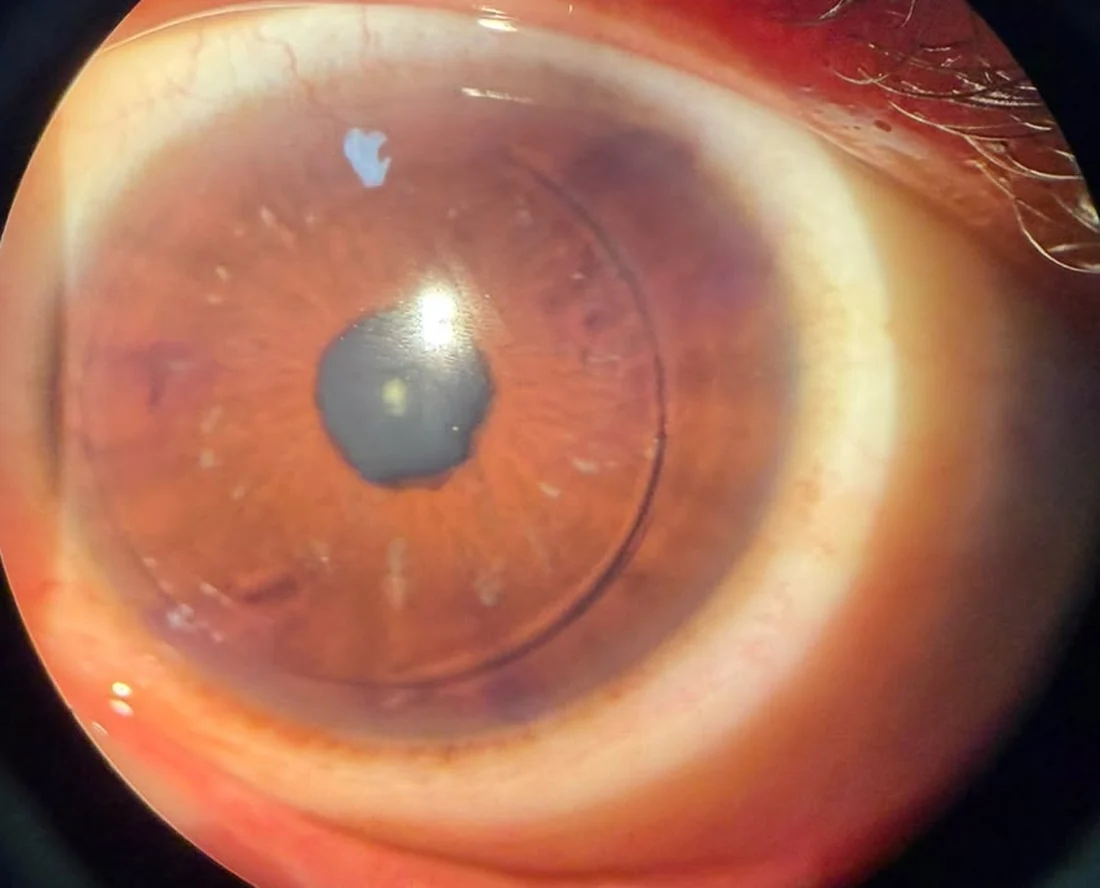
The rest of the examination was essentially normal. Full ocular movements and the patient in orthophoria, apart from his nystagmus. Three minor findings are worth naming, because they are part of a complete workup. A small inferior area of atonic pupil in the left eye with discreet uveal ectropion in the same sector, inferior punctate epithelial erosions, and a cup-to-disc ratio of 0.65 in both eyes. That figure is always read against the size of the optic disc, because a large disc can have a wide cup without being pathological, so on its own it diagnoses nothing. It does call for following the optic nerve over time, with visual fields and OCT of the nerve fibre layer.
What a lens cannot correct
Here is the part that explains the case.
A lens corrects two things, sphere and cylinder. Those describe the simplest distortions of the optical system, which is why most people see well with their prescription in place. But a cornea can also carry more complex irregularities, known in optics as higher order aberrations. They are not described by those two numbers and no conventional lens compensates them.
The study with the Topolyzer, which measures the shape of the cornea point by point, showed that this patient had significant higher order aberrations. And that raises the question that organises the whole case. If part of his poor vision came from the shape of the cornea rather than from the brain, that part was treatable.
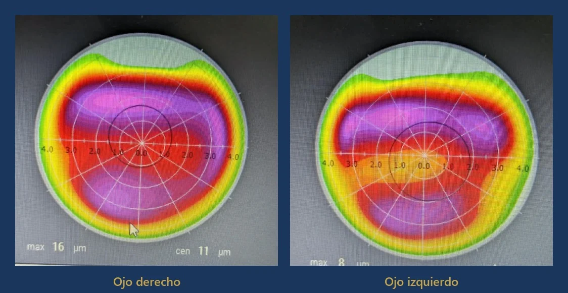
That asymmetry is the visual proof of the point. Two eyes with nearly identical prescriptions can need very different treatments, because what separates them is not in the refraction but in the shape.
What was done
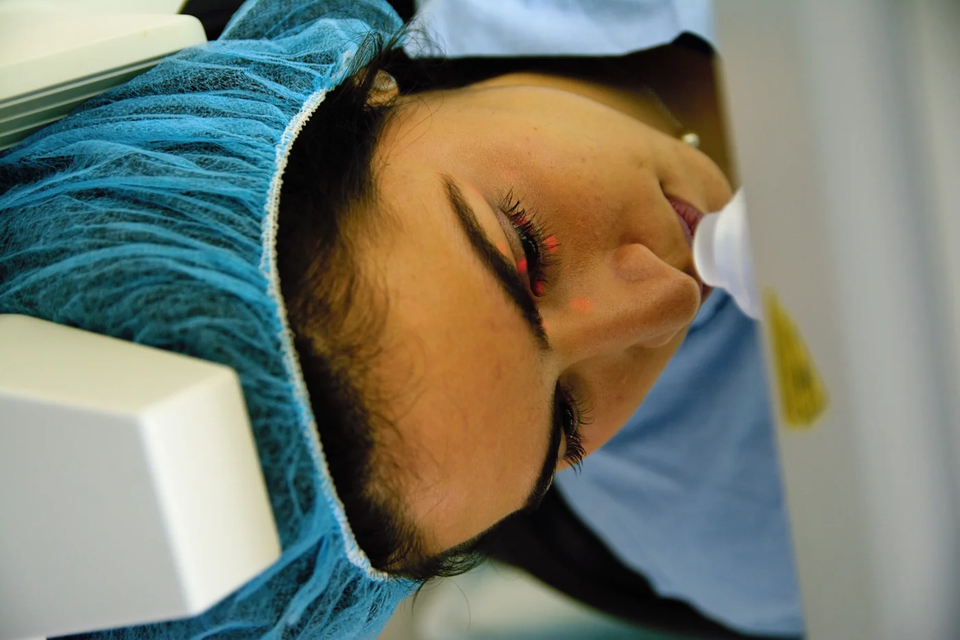
On 10 September 2026 a bilateral topography-guided FEMTO-LASIK was performed, without complications.
The flaps were created with the VisuMax 500 femtosecond laser. The ablation was carried out with the WaveLight EX500 excimer, incorporating the Topolyzer data to generate an individualised treatment for each cornea rather than applying the prescription alone.
One technical detail deserves explaining, because it is not obvious. When the laser regularises an irregular cornea, that regularisation by itself changes the eye’s prescription. Feed in the consulting-room refraction unchanged and the result overshoots or falls short. For that reason the treatment refraction was adjusted using the team’s own calculation protocol, to compensate for the changes induced by the topographic component of the ablation.
Before surgery he was told clearly what it could and could not do. It could correct the refractive error and improve the optical quality of the cornea. It could not guarantee visual acuity beyond whatever limit the amblyopia imposed.
The result, and why it is surprising
At the next-day visit, uncorrected:
| Before, best corrected | Day 1, uncorrected | |
|---|---|---|
| Right eye | 20/40+1 | 20/30 |
| Left eye | 20/40−1 | 20/25 |
About one line of improvement in the right eye and two in the left. And the comparison is not against an ideal chart, it is against himself: he is seeing, with nothing in front of his eyes, better than he saw with the best correction that could be placed there.
Reading the case
The result suggests that his preoperative limitation was not determined exclusively by refractive amblyopia. An optical component, related to the irregularity of the cornea and to the higher order aberrations, appears to have played a clinically relevant role.
Put another way. The diagnosis of amblyopia was correct, but amblyopia did not account for the whole deficit. Underneath it lay an optical part that spectacle correction could not reach, because spectacles do not correct that kind of distortion. Regularising the surface recovered it.
This is not an isolated phenomenon. Several published series describe a proportion of amblyopic eyes gaining corrected acuity after refractive surgery. In the most robust of them, which compared each amblyopic eye against the healthy fellow eye of the same patient, 27.9% of amblyopic eyes gained two or more lines of corrected acuity, against 6.2% of the fellow eyes (Liu, Schallhorn et al., Journal of Refractive Surgery, 2020). A gap that wide between the two eyes of the same person is hard to explain by chance or by familiarity with the chart. These are retrospective studies, and no randomised trials confirm them.
“This case shows the importance of distinguishing between a true visual limitation caused exclusively by amblyopia and a reduction in vision in which potentially correctable corneal optical aberrations coexist.”
Dr. Juan F. Batlle Logroño, ophthalmologist subspecialised in cornea and refractive surgery
The distinction has practical consequences. An adult with amblyopia is usually told there is nothing to be done, and for the strictly amblyopic part that is true. The question this case invites is a different one: how much of what he cannot see is amblyopia, and how much is uncorrected optics? That is answered by measuring the cornea, not by assuming.
What this case does not prove
The limits are worth stating out loud, because that is what separates a clinical report from an advertisement.
This is one patient and one postoperative day. First-day vision is not final vision in any refractive surgery. It can keep improving for weeks and can also regress while the cornea heals. Final acuity will be judged over the follow-up.
The amblyopia was not reversed. Nothing here indicates that the visual pathway changed. What changed is the quality of the image reaching that pathway.
The nystagmus was not corrected either, nor was that the intention. It is still there and will remain.
And there is no literature on this. A PubMed search for topography-guided treatment in amblyopic eyes returns no published series. What exists is evidence on each side separately. Refractive surgery in amblyopic eyes on one hand, topography-guided treatment in normal eyes on the other. This case falls in the gap between them, and is published as what it is, an isolated observation where no series exists, rather than the application of something already established.
Nor is it being claimed that the aberrations went down. Sustaining that would require postoperative aberrometry compared against the preoperative measurement, and there is none here. It is also worth knowing that the ablation itself induces new aberrations, and that several studies measure higher total aberrations after surgery than before, even with a topography-guided profile. What this case documents is an improvement in acuity, not a measured reduction in aberrations.
Nor does it mean that any adult with amblyopia will have this result. Several conditions coincided here. A cornea with measurable and treatable aberrations, sufficient thickness, a stable prescription, and a patient who understood that the goal was optical quality rather than a number. With any of those missing, the conversation is different.
What this case does change in the clinic
Being told you have amblyopia does not close the question. A workup that separates the two things closes it.
If you have carried a high astigmatism since childhood and have been told your vision cannot improve, the reasonable step is not to look for someone who will promise otherwise, but to ask for the shape of your cornea to be measured and for someone to explain how much of your deficit is optical. The answer may still be that there is no margin. And there may be a part nobody had looked at.
One last thing, which is not clinical
This patient is over two metres tall, played basketball and is today an avid golfer. Before surgery he could not follow the ball’s flight with his eyes, so he played using an artificial intelligence app on his phone that tracked the ball so he could find it.
His expectation, twenty-four hours after surgery, was to stop needing it.
That is the kind of thing that does not appear on a visual acuity chart and that, for the person living it, is the result.
Medical note. This article is informative and educational in purpose. It describes an individual clinical case and does not constitute a treatment recommendation, nor does it replace evaluation by an ophthalmologist. The outcomes of refractive surgery depend on the characteristics of each eye and are not transferable from one patient to another. The case is published anonymously and with the patient’s consent.
About the author. Dr. Juan F. Batlle Logroño is an ophthalmologist subspecialised in cornea and refractive surgery. He read Medicine at Tulane University School of Medicine, completed his residency in Ophthalmology at Tulane and his fellowship in Cornea and External Diseases at the Bascom Palmer Eye Institute, University of Miami. He practises in Santo Domingo, Dominican Republic.
Sources
- American Academy of Ophthalmology. Amblyopia Preferred Practice Pattern. Ophthalmology, 2022. Full text
- Harvey EM. Development and treatment of astigmatism-related amblyopia. Optometry and Vision Science, 2009. Abstract
- Liu X, Schallhorn SC et al. Three-month outcomes of laser vision correction for myopia and hyperopia in adults with amblyopia. Journal of Refractive Surgery, 2020. Abstract
- Felius J, Muhanna ZA. Visual deprivation and foveation characteristics both underlie visual acuity deficits in idiopathic infantile nystagmus. Investigative Ophthalmology and Visual Science, 2013. Abstract
- İpek ŞC, Utine CA. Topography-guided excimer laser ablation in refractive surgery. Frontiers in Ophthalmology, 2024. Full text