There is one figure that changes the way you look at what happens in a delivery room. A single placenta donated after a scheduled cesarean section can yield up to ~30 amniotic membrane grafts, tissue that would otherwise be discarded as medical waste (PMC4094946, 2014). That tissue, properly processed, becomes one of the most useful tools we have to repair the surface of the eye when the cornea cannot heal on its own.
The amniotic membrane is not a drug nor a "stem cell" transplant, although those mix-ups are very common. It is the innermost layer of the placenta, and once processed it works as a biological patch: a scaffold on which the corneal epithelium grows back, loaded with factors that calm inflammation and curb uncontrolled scarring. Its use in ophthalmology is neither new nor experimental. It was first documented in 1940 and became established again in ocular surface surgery during the 1990s. In that early stage, at Bascom Palmer, my father, Dr. Juan F. Batlle Pichardo, presented one of the first case series with amniotic membrane on the ocular surface. It is part of the history I inherited and continue to apply in my practice.
I want to explain to you, without unnecessary jargon, what the amniotic membrane is, why it helps the ocular surface heal and in which cases it has a clear role and in which it does not. And, because it is part of my daily work, where this tissue comes from and why donation matters so much.
What the amniotic membrane is and why it works
The amniotic membrane is the layer in direct contact with the fetus inside the placenta. It has an epithelium, a thick basement membrane and an avascular stroma (without blood vessels). What is interesting for us is that the collagen composition of that basement membrane is similar to that of the cornea and conjunctiva, which makes it a substrate almost tailor-made for the ocular surface (Annals of Eye Science, 2021).
Once processed, the membrane is essentially acellular. It does not act because it "transplants cells," but through two pathways. Its matrix (collagen types IV and VII, laminin, fibronectin) gives the epithelium an ordered surface to adhere to and migrate over, and it releases a cocktail of growth factors, among them EGF, KGF, HGF and bFGF, that stimulate repair (PMC4863510, 2016).
In my experience, the clearest way to explain to a patient what the membrane does is to talk about its four properties, the ones we usually call "the four antis":
- Pro-epithelialization. It serves as a scaffold for epithelial cells to grow, differentiate and adhere, and it prevents their premature death.
- Anti-inflammatory. It contains molecules that switch off inflammation, traps inflammatory cells and blocks enzymes that damage tissue.
- Antifibrotic. It suppresses TGF-β signaling, which is the pathway that triggers excessive scarring. In practice, that means less opaque scarring on the cornea.
- Antiangiogenic. It releases factors that curb the formation of new blood vessels, vessels that should not exist in a healthy cornea because they reduce transparency.
That combination is what explains why a single tissue serves such different problems. And there is a detail that reassures almost every patient when I explain it: the membrane lacks the HLA-A, -B and -DR antigens, the markers that trigger rejection. That is why, although it is technically donor tissue, rejection is very infrequent (AAO EyeNet).
How it reaches the eye: with or without sutures
There are several ways to preserve the membrane. The cryopreserved form is kept at −80 °C and retains its proteins and growth factors very well. The dehydrated form is stored at room temperature, lasts longer and is easier to distribute (StatPearls/NCBI, 2023). There is also alcohol preservation, a more economical method that broadens access to the tissue. At the Dominican Republic Cornea Bank we use this approach precisely so that cost is not a barrier and more patients can benefit.
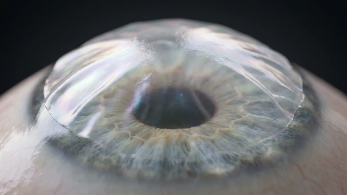
In the operating room, the membrane can be sutured onto the cornea as a graft when the case warrants it. But there is also the sutureless format, a ring-shaped device that holds the membrane and is placed in the office. In these cases it usually dissolves on its own in about 3 to 7 days, faster if there is a lot of inflammation, and FDA regulation establishes that it should not remain more than 29 days in the eye (PMC10573988, 2023). For the patient, this means that the treatment is often outpatient and well tolerated.
What it really helps with (and what it does not)
Here it is worth being honest, because the amniotic membrane is not a universal solution. It is one more option, very valuable in certain scenarios and modest in others. I always say it in consultation: what gives a treatment credibility is knowing where it delivers and where it does not.
Where its role is clearest is in the ocular surface that cannot close. In persistent epithelial defects and ulcers that do not heal with medical treatment, the membrane achieves complete closure in 58 to 91% of cases depending on the series, with an overall success of 74.4% in a multicenter study, and around 70% in corneas coming from a prior transplant (PMC8348886, 2021; PubMed 18535612, 2008). That is the situation where the membrane earns its place: it is a rescue procedure when conservative treatment has already failed.
In chemical burns the picture is more nuanced, and this nuance matters. In moderate burns (grades II–III), a clinical trial showed that the membrane accelerates epithelialization, with 15.9 days versus 28.9 days with medical treatment alone, and a better final visual acuity (Tandon, 2011; Cochrane, 2022). But in severe grade IV burns, the highest-level evidence, a 2022 Cochrane review, shows no benefit: epithelialization was 75.8 versus 72.6 days, with no significant difference (Eslani, 2019). In other words, the membrane helps when the damage is moderate, but it is not the solution when the burn has completely devastated the surface.
In severe dry eye, which is an enormous and very common problem, the membrane has a regenerative role that I find fascinating. A clinical trial found that three months after a placement, corneal nerve density increased by around 54% and corneal sensitivity by 72% (PMC5574308, 2017). It is not just covering the eye, it is helping the cornea recover its innervation.
There are two other situations where it is worth spelling out the limits frankly:
- Pterygium. Here the membrane can be used, but the highest-level studies are clear: conjunctival autograft (using the patient's own tissue) prevents recurrence better. A Cochrane review showed that the autograft reduces recurrence 47% more than the membrane (RR 0.53, 95% CI 0.33–0.85). When the goal is for the pterygium not to come back, the autograft is usually the better option.
- Neurotrophic keratopathy. The membrane helps the ulcer heal, in 76 to 86% of cases, but recurrence is around 46% at twelve months (PMC8850233, 2021). The reason is important: the membrane closes the wound, but it does not correct the underlying denervation, which is the real cause. It treats the consequence, not the origin.
The membrane also works as support in ocular surface reconstruction surgeries, such as limbal stem cell transplantation, where it serves as a base for the new epithelium to establish itself. In all these scenarios the rule I repeat is the same: the membrane is a tool for visual rehabilitation and surface rescue, not a substitute for treating the underlying disease.
A confusion worth clearing up: these are not stem cells
The question I hear most about this topic mixes up two different things. The amniotic membrane as a patch or graft is a legitimate treatment, with decades of clinical backing, and it is not the same as the "amniotic fluid drops" promoted online as a miraculous regenerative therapy. The FDA issued a public safety notification against those drops in 2023, which are not approved (FDA, 2023).
The distinction is simple: the membrane works through its matrix and its growth factors, not because it contains living cells that are transplanted. When someone offers you a treatment "with stem cells from the cord or amniotic fluid" for the eye, it is worth asking exactly what they are putting in and under what backing. Part of my job is to separate well-used biology from dubious marketing.
How this membrane was identified: a story that began in my family
That we use amniotic membrane so naturally today hides a story that is close to me, because one piece of it was solved here, in the Dominican Republic, and my own family was behind it.
In the late 1980s, in the former Soviet Union, Professor Muldachev, of the University of Ufa, operated with a tissue that healed the surface of the eye but kept its origin a secret. He simply called it allotransplant. That tissue reached Venezuela, and from there Dr. Carlino González brought a sample to Santo Domingo, into the hands of my father, Dr. Juan F. Batlle Pichardo, with an unanswered question: what was it really?
My father did not solve it alone. He gave that sample to my maternal grandfather, Dr. Miguel Ángel Logroño, who was a pathologist, and in order to compare, he separated the amniotic membrane from a local placenta himself and sent it to biopsy as well. My grandfather studied both tissues under the microscope, compared them, and confirmed that they were exactly the same: amniotic membrane. In 1992, my father and Dr. Francisco Perdomo presented that finding at the American Academy of Ophthalmology meeting and at the Bascom Palmer Eye Institute. By then, that membrane had already helped 23 patients with ulcers, burns, and other difficult ocular surface conditions. The work was later documented in a chapter on the history of amniotic membrane in pterygium surgery.
I tell this story for two reasons. The first, because it explains where the technique I can offer a patient today comes from. The second, because there is a thread running through it: a grandfather pathologist who confirmed the tissue under the microscope, a father surgeon who solved the mystery, and me, still using that membrane every week to help heal eyes. The same calling, across three generations.
Where this tissue comes from: the journey of donation
This is the part that touches me most, because it connects with the place where I work. The amniotic membrane is obtained from placentas donated after a scheduled cesarean section, never from a vaginal delivery, because the vaginal canal contaminates the tissue with bacteria. The scheduled cesarean offers a sterile and controlled environment (PMC11235369, 2023).
The process is rigorous. The donor mother signs informed consent and undergoes complete serological testing: HIV, hepatitis B and C, syphilis, HTLV and CMV. The tissue remains in quarantine and the tests are repeated to cover the window in which a recent infection has not yet appeared in the analyses, and it is approved only if all come back negative. It is worth emphasizing: there are no published reports of disease transmission from ocular amniotic membrane (AAO EyeNet; StatPearls/NCBI, 2023).
Here I return to the figure from the beginning, because it sums up everything I want to convey. A single placenta, tissue that normally ends up in the trash as medical waste, can become up to ~30 grafts that restore vision to dozens of people (PMC4094946, 2014).
Why having a tissue bank in the Dominican Republic matters
Access to ocular tissue is a serious global problem. In the world there are about 5.5 million people with corneal blindness in both eyes, and the supply of tissue is dramatically insufficient: there is barely 1 cornea available for every 70 that are needed, and around 53% of the world's population has no access to ocular tissue (Gain et al., JAMA Ophthalmology, 2016). Latin America donates well below its demand.
In that context, having a local tissue bank is not a luxury, it is infrastructure that brings treatments closer to Dominican patients and reduces dependence on imported tissue. The Dominican Republic Cornea Bank was founded in 1987 (my father, Dr. Juan F. Batlle Pichardo, was one of its founders, along with Freddy Beras Goico and Dr. Luis Cuello Mainardi) and has been a member of the Pan-American Association of Eye Banks since 1989. Today I am Medical Director of that bank, and part of my work is ensuring that the necessary tissue is available when a patient needs it.
Tissue logistics is, very often, what separates a possible intervention from an impossible one. I experienced it firsthand in the structural rescue of a keratoprosthesis with corneal melt, where rapid access to donor tissue was the difference between saving the eye and losing it. Something similar happens with the amniotic membrane: having it available makes it possible to offer surface rescues within timeframes that otherwise would not be met.
The availability of amniotic membrane varies between centers. In some cases it is obtained from local tissue banks and in others imported processed products are used. What matters for the patient is that, when the surface of the eye needs this biological support, there is a way to obtain it in time and with the appropriate quality controls.
What a patient can expect
In practice, the membrane is usually well tolerated. With the sutureless format the patient may feel the ring, although it generally does not hurt, and may notice temporary blurry vision while the membrane covers the eye, until it dissolves on its own within a few days. When it is sutured as a graft, the postoperative course depends on the underlying case.
As with everything in cornea, this depends on the case. Before proposing an amniotic membrane, the protocol includes studying the ocular surface, identifying the underlying cause and deciding whether the membrane is the right tool or only a patch that delays the true solution. If you want to learn how we approach ocular surface and dry eye problems, you can review our services.
Frequently asked questions
Is the amniotic membrane a transplant? Will my body reject it?
Technically it is a donor tissue graft, but the membrane is immunologically privileged: it lacks the markers that trigger rejection (HLA-A, -B, -DR). That is why rejection is very rare, unlike what happens with other transplants.
Are these stem cells?
No. The amniotic membrane for the eye is essentially acellular and works through its matrix and its growth factors, not through stem cells. It should not be confused with the "amniotic fluid drops" sold online, which the FDA warned in 2023 are not approved.
Where does it come from and is it safe?
It comes from placentas donated by healthy mothers after a scheduled cesarean section, with informed consent, complete screening for HIV, hepatitis and syphilis, and tissue quarantine. There are no published reports of disease transmission from ocular amniotic membrane.
Does it hurt and how long does it stay in place?
It is generally well tolerated. With a sutureless device you may feel the ring, but it does not usually hurt, and the membrane dissolves on its own in about 3 to 7 days, faster if there is a lot of inflammation.
Does it work for any eye problem?
It is not a universal solution. It has a clear role in non-healing ulcers and defects, moderate burns, severe dry eye and as support in ocular surface reconstruction surgery. In pterygium, conjunctival autograft prevents recurrence better, and in neurotrophic keratopathy it heals but does not correct the underlying cause.
Disclaimer
This content is for educational and informational purposes. It does not replace professional ophthalmological consultation, diagnosis or treatment. The decision to use an amniotic membrane and the choice of technique should be made with a cornea specialist after an individual evaluation. The efficacy figures cited come from published studies and describe probabilities of response, not guarantees; results may vary according to each person's clinical conditions.
References
- Walkden A. Amniotic Membrane Transplantation in Ophthalmology: An Updated Perspective. Clin Ophthalmol / PMC8966384. 2022.
- Liu J, et al. New clinical application of amniotic membrane transplantation for ocular surface disease. PMC4863510. 2016.
- Jirsova K, Jones GLA. Amniotic membrane in ophthalmology: properties, preparation, storage and indications. Cell Tissue Bank / Springer. 2017.
- Tandon R, et al. Amniotic membrane transplantation as an adjunct to medical therapy in acute ocular burns. Br J Ophthalmol. 2011. PMID: 20675729.
- Clare G, et al. Amniotic membrane transplantation for acute ocular burns. Cochrane Database Syst Rev. 2022. PMC9435439.
- Eslani M, et al. Amniotic membrane transplantation in acute severe ocular chemical injury (ECA). 2019. PMID: 30419194.
- John T, et al. Corneal nerve regeneration after self-retained cryopreserved amniotic membrane in dry eye disease (ECA). PMC5574308. 2017.
- Sutureless self-retained amniotic membrane for ocular surface disorders. PMC10573988. 2023.
- Clearfield E, et al. Conjunctival autograft for pterygium. Cochrane Database Syst Rev. 2016/2017. PMC5610642.
- Mun Y, et al. Cenegermin vs amniotic membrane in neurotrophic keratopathy. PMC8850233. 2021.
- Preparation of human amniotic membrane for transplantation. PMC11235369. 2023.
- Human amniotic membrane: different modalities of use and yield. PMC4094946. 2014.
- Gain P, et al. Global Survey of Corneal Transplantation and Eye Banking. JAMA Ophthalmol. 2016.
- Flaxman SR, et al. Global trends in corneal opacity blindness. Ophthalmology. 2023.
- U.S. FDA. Public Safety Notification on Amniotic Fluid Eyedrops. 2023.
Last updated: June 23, 2026