Keratoconus affects approximately 1 in 2,000 people according to data from the National Eye Institute, and that figure likely underestimates the reality because many mild cases go undiagnosed. It is a condition in which the cornea loses its spherical shape, progressively thins, and takes on a conical geometry that significantly distorts vision. For decades, the only response to advanced keratoconus was a corneal transplant. That changed with corneal crosslinking.
In 2007, CCCRP brought to the Dominican Republic the first ultraviolet light device for corneal crosslinking with riboflavin. We were the first center in the country to offer this treatment. Since then, I have been able to observe how crosslinking has changed the prognosis of hundreds of patients who previously had a transplant as their only prospect. That accumulated experience of nearly two decades is the foundation of what I share in this article.
What crosslinking does and why it works
The principle is simpler than it sounds. The collagen fibers that form the cornea are linked to each other through biochemical bonds. In keratoconus, these bonds are insufficient, which allows the cornea to deform under intraocular pressure. Corneal crosslinking (CXL) creates new bonds between these fibers through a photochemical reaction: riboflavin (vitamin B2) is applied to the cornea and then irradiated with ultraviolet light type A (UV-A). The combination generates free radicals that induce the formation of additional covalent bridges between collagen molecules.
The result is a cornea that is more rigid and more resistant to deformation.
This is not about reversing the damage that already exists. The goal of crosslinking is to halt or slow the progression of keratoconus by increasing the biomechanical rigidity of the corneal tissue. In a long-term study published in Current Eye Research (2022), 99.2% of eyes treated with CXL remained stable with no signs of progression after three years of follow-up. Those figures back up what I see in daily clinical practice.
Who is a candidate for corneal crosslinking?
Not every patient with keratoconus needs crosslinking, and not all are ideal candidates. The most favorable profile is a young patient (between 18 and 44 years old) in the early or intermediate stages of the disease, where the cornea still has sufficient thickness and progression is documented.
The criteria I evaluate include:
- Confirmed diagnosis of progressive keratoconus or post-surgical corneal ectasia
- Minimum corneal thickness of 400 microns (measured with Pentacam)
- Documented progression: increase in maximum keratometry, change in topography, or deterioration of corrected visual acuity
- Absence of significant corneal scarring
- No active ocular infection
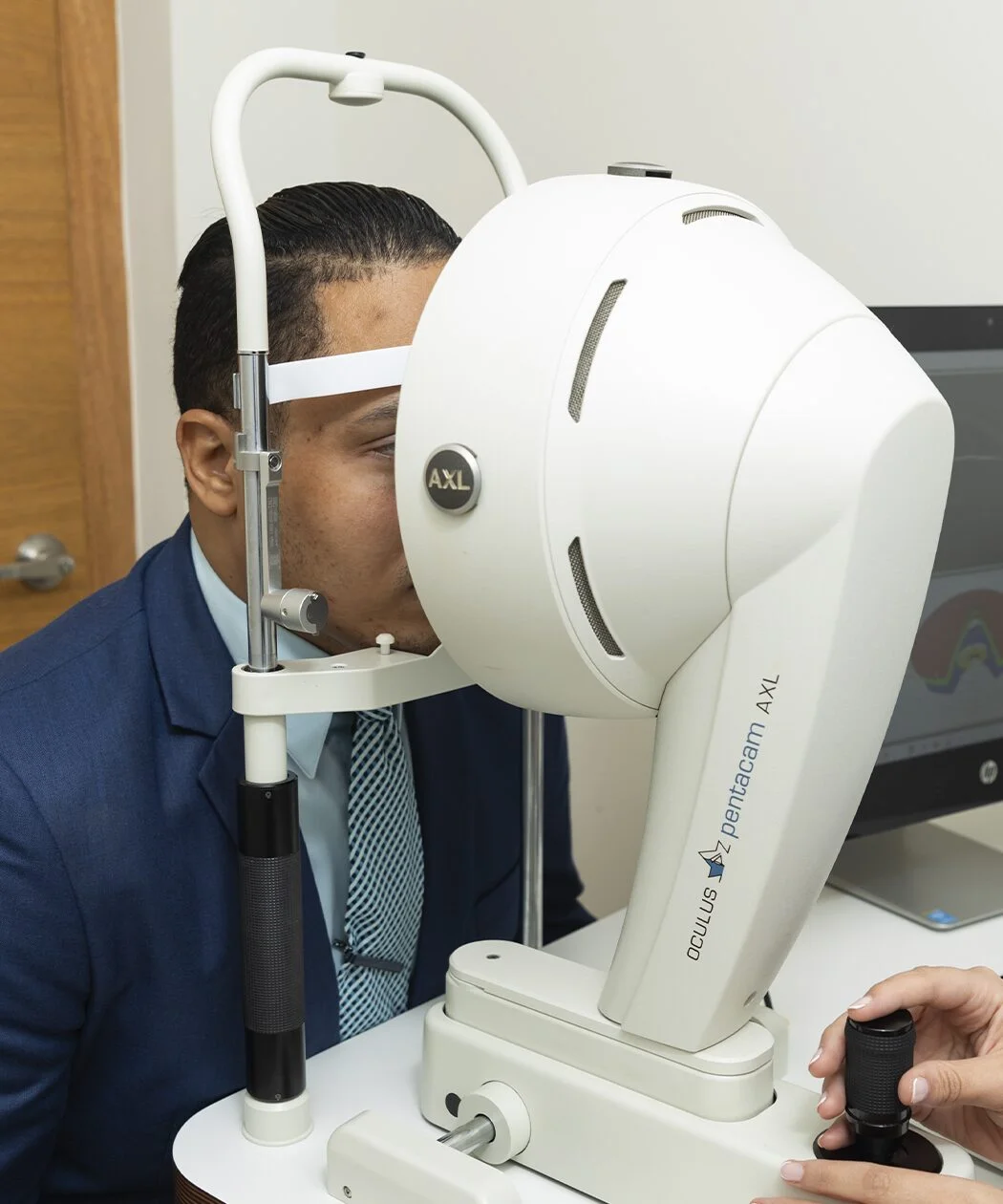
The pre-op evaluation is meticulous. At CCCRP we use Pentacam to map corneal elevation and full thickness, anterior segment OCT to evaluate the layers of the cornea in detail, and specular microscopy to count endothelial cells. Each case of keratoconus is different, and the decision to perform crosslinking must be based on objective evidence of progression, not solely on the diagnosis.
Conditions treated with CXL
Crosslinking is not exclusive to keratoconus. It is used in three main scenarios:
- Progressive keratoconus: the most frequent and best-studied indication.
- Post-surgical corneal ectasia: weakening of the cornea that can appear after refractive laser surgery (LASIK, PRK), where the cornea did not have enough biomechanical reserve.
- Pellucid marginal degeneration: a form of peripheral corneal ectasia, less common but equally treatable with CXL.
In all cases, the mechanism is the same: strengthening a cornea that has lost its structural integrity.
How the procedure works, step by step
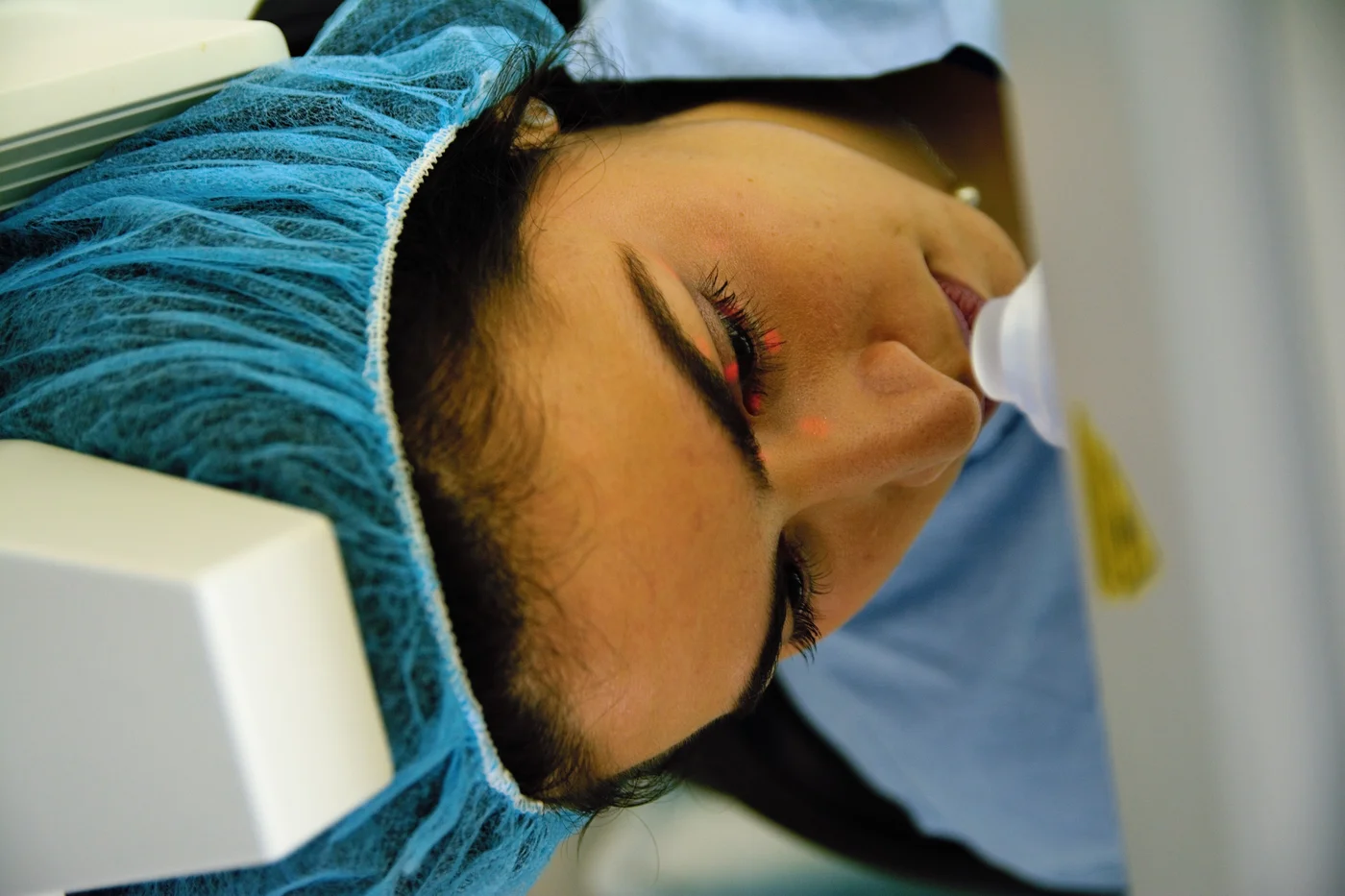
Corneal crosslinking is an outpatient procedure that lasts approximately one hour. It is performed under topical anesthesia (drops), which means the patient feels no pain during the intervention.
The standard protocol (known as the Dresden protocol) follows these steps:
- The central corneal epithelium is removed to allow riboflavin to penetrate the stroma.
- 0.1% riboflavin drops are applied to the cornea for 30 minutes, verifying tissue saturation.
- The cornea is irradiated with UV-A light at 3 mW/cm² for 30 minutes, while riboflavin application continues.
- A therapeutic contact lens is placed and remains until the epithelium regenerates (between 3 and 5 days).
Full recovery takes two to four weeks. The first few days can be uncomfortable: tearing, light sensitivity, and blurred vision are normal while the epithelium heals. Vision stabilizes gradually over the following months.
Standard CXL vs. accelerated CXL
There is a variant of the procedure called accelerated crosslinking, which uses higher UV light intensity for a shorter time. The idea is to reduce the total duration of treatment.
| Feature | Standard CXL (Dresden) | Accelerated CXL |
|---|---|---|
| Irradiation duration | 30 minutes | 5 to 10 minutes |
| UV-A intensity | 3 mW/cm² | 9 to 30 mW/cm² |
| Total time | ~60 minutes | ~30 to 40 minutes |
| Riboflavin penetration | High | Variable depending on protocol |
| Long-term evidence | Extensive (more than 15 years) | Growing, lower volume |
| Demonstrated efficacy | Reference standard | Comparable in one-year studies |
In my practice, I select the protocol based on the individual case. Standard CXL remains my first choice for most patients due to the strength of the supporting evidence.
Real benefits of crosslinking
I want to be direct about what crosslinking can and cannot do:
- It can halt or slow the progression of keratoconus in the vast majority of treated cases.
- It can delay or prevent the need for a corneal transplant, which is a far more invasive procedure with risk of rejection and years of recovery.
- It can improve long-term visual stability, allowing the patient to continue wearing lenses with a more predictable prescription.
- It does not cure keratoconus. The existing deformation is not reversed with CXL. Some patients experience mild corneal flattening after treatment, but this is not the norm.
For a young patient with progressive keratoconus, crosslinking can mean the difference between maintaining functional vision for decades or needing a transplant before age 40.
A milestone for Dominican ophthalmology
When in 2007 we installed at CCCRP the first ultraviolet light device for crosslinking in the Dominican Republic, many colleagues were still unfamiliar with the technique. The evidence from the Wollensak, Spoerl, and Iomdina group had been published only four years earlier. Committing to CXL at that moment was a decision based on the literature and on the conviction that our patients deserved access to the same advances available at international reference centers.
Nearly two decades later, crosslinking is the standard of care for progressive keratoconus worldwide.
Frequently asked questions
Does corneal crosslinking hurt?
The procedure is performed with topical anesthesia (drops), so no pain is felt during the intervention. In the days that follow, it is normal to experience discomfort, tearing, and light sensitivity while the corneal epithelium regenerates. These symptoms are managed with medication and usually resolve within a week.
At what age is crosslinking recommended?
The usual range is between 18 and 44 years old. Keratoconus tends to progress more aggressively in young patients, which makes early detection and timely treatment especially relevant in this population.
Might I need another treatment after crosslinking?
Yes. Crosslinking halts progression, but it does not correct the existing refractive error. Many patients continue using specialized contact lenses after the procedure. In some cases, complementary treatments such as intrastromal rings or intraocular lenses can be combined.
How long does visual recovery take?
Vision is usually blurred during the first two weeks while the epithelium heals. Full visual stabilization can take between one and three months.
Can crosslinking be repeated?
In theory yes, and there are reports of retreatment in cases where progression is documented after the first CXL. However, this is uncommon. The decision to retreat requires careful evaluation of the residual corneal thickness and the state of the endothelium.
References
- Wollensak, G., Spoerl, E., & Seiler, T. (2003). Riboflavin/ultraviolet-A-induced collagen crosslinking for the treatment of keratoconus. American Journal of Ophthalmology, 135(5), 620-627.
- Raiskup, F., et al. (2022). Long-term outcome of corneal collagen crosslinking with riboflavin and UV-A irradiation for keratoconus. Current Eye Research, 47(11), 1543-1550.
- Gustafsson, J., et al. (2025). Early findings in a randomised controlled trial on crosslinking protocols. Acta Ophthalmologica.