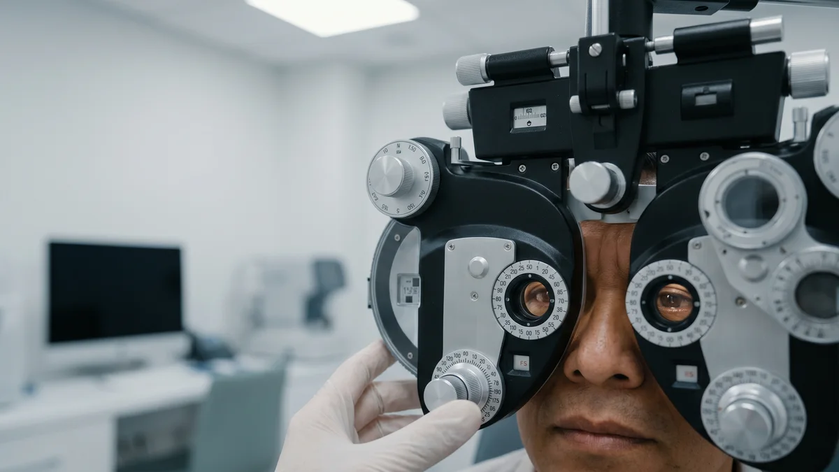
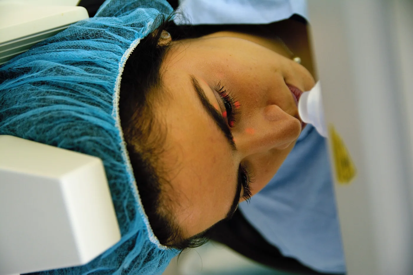
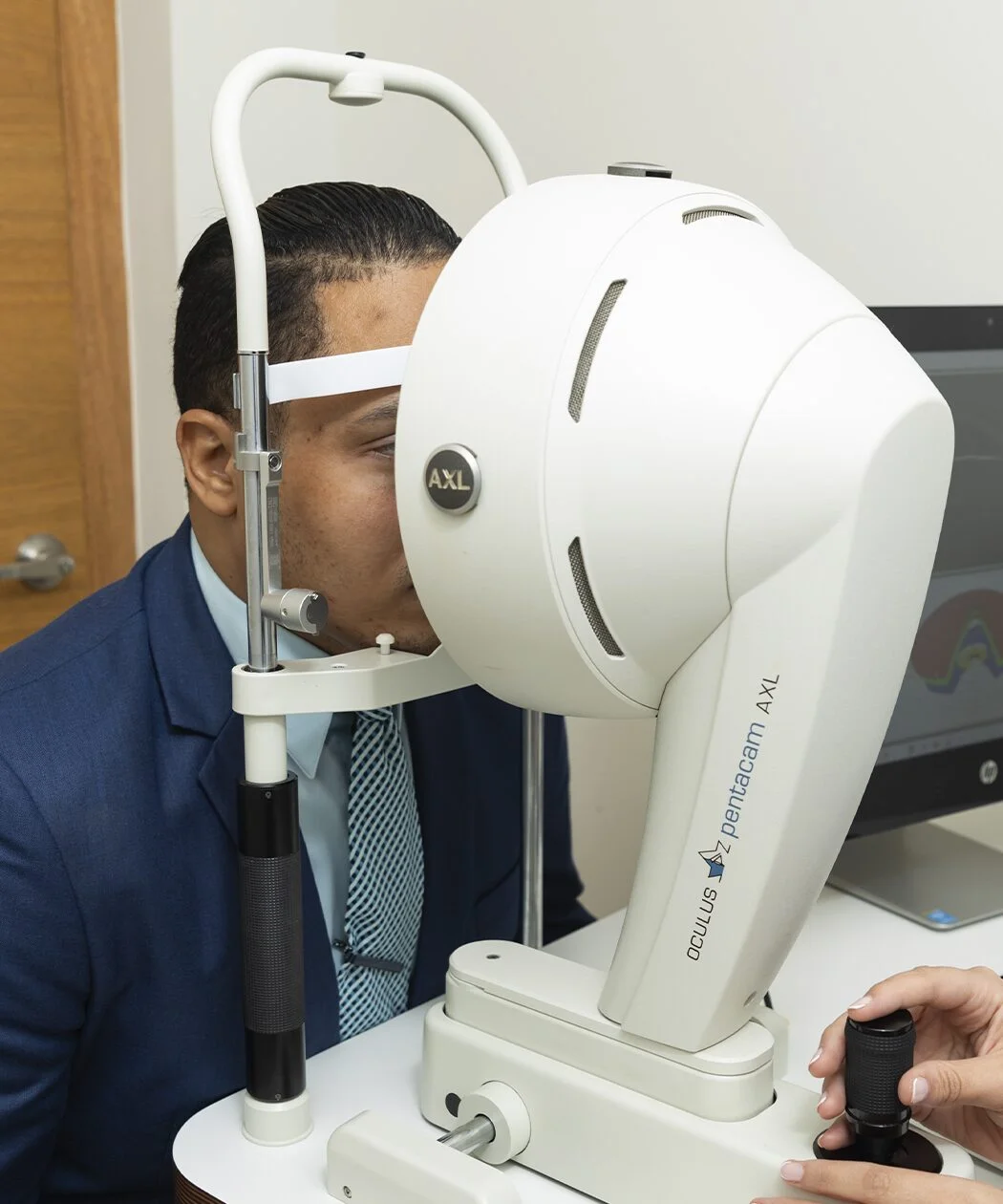
A patient from the United States arrives in Santo Domingo for hyperopia surgery. Without glasses she saw 20/400 in each eye, which means not being able to function without them. With her prescription in place, +5.00 dioptres in one eye and +4.25 in the other, she saw 20/20. Her prescription had been unchanged for two years.
She was a textbook candidate for a phakic lens, the one implanted inside the eye without touching the cornea and without removing anything. But she had two problems, and neither was her prescription.
The first is administrative and explains the trip. That lens, in its hyperopic version, is not approved in the United States. The Food and Drug Administration (FDA) approved it for myopia in December 2005, for myopic astigmatism in September 2018, approved the current design in March 2022 and in November 2025 widened the indicated age up to 60. All of those indications are for myopia. For hyperopia there is no approved version there, although the lens exists and is implanted in the markets governed by the European marking.
The second problem was anatomical, and it is the one that makes the story. It had to do with the size of the space that lens would have to go into.
The figure that disqualified her
Her anterior chamber, the space between the cornea and the crystalline lens where the implant has to fit, measured 2.82 millimetres in one eye and 2.84 in the other. The rule we have worked with for years asks for a minimum of 3.0 millimetres, measured from the corneal endothelium. It appears in the instructions for use of the hyperopic lens itself, and the same threshold appears in the US labelling of the lens for myopia. It is there for a clear reason. A lens forced into a space too small compresses the iris and the ciliary body, the angle through which the eye drains its fluid narrows, and pressure can rise and angle-closure glaucoma appear.
With that figure in hand, the short answer is that she is not a candidate. It is the answer she had already been given twice.
What I argue is that the short answer has grown old. The threshold is still there, but it is no longer the only thing available to look at.
“With the Guru you can now push the limits that had been set for us before. If in hyperopia we were told that nobody qualified with a chamber under 3 millimetres, today you can predict how the iridocorneal angle and the lens vault will end up and, if they fall in the safe range, you can still implant even below the previous limit.”
Dr. Juan F. Batlle Logroño, ophthalmologist subspecialised in cornea and refractive surgery
The Guru is an AI-assisted sizing program that, rather than giving a size, predicts for every available size how the vault and the angle will turn out. The three-millimetre threshold was a safety rule built when none of that could be predicted. If the outcome can now be estimated eye by eye, the question stops being whether the figure clears the bar and becomes what will happen in this particular eye.
With one condition that I add, and it is half the matter. The prediction is worth what the measurements fed into it are worth, and the ones I feed it come from ultrasound.
What was planned from outside
The ophthalmologist who referred her sent the complete workup in advance, so that she would make one trip instead of three. With those measurements, the manufacturer’s calculator recommended a lens size of 12.6 millimetres for both eyes.
That recommendation comes, among other things, from the visible diameter of the eye across the coloured part, what in clinic is called white to white. It is an external measurement, and the lens sits behind the iris, in a sulcus that cannot be seen from outside.
So a second pair of lenses, 12.1 millimetres, was ordered in case the inside measurements did not match the outside ones. Both pairs were in Santo Domingo before she landed. That precaution turned out to be the most important decision of the case.
The V pattern, explained
The day before surgery she had ultrasound biomicroscopy, which passes through the iris and draws what lies behind. It is the only way to measure directly the sulcus where the lens will rest, instead of inferring it from outside. And it showed that this sulcus was considerably smaller than the external measurement suggested.
That discrepancy is described and measured in the literature, which calls it the difference between sulcus-to-sulcus and white-to-white. I explain it in clinic with an image that lands straight away.
“When you look at the eye in a sagittal section, the distance from sclera to sclera, white to white, at the limbus is greater than the sulcus-to-sulcus distance, which is the walls of the eye just behind the iris. If you drew a line on each side through each distance and joined it in the middle to the line from the opposite side, it would form a V.”
Dr. Juan F. Batlle Logroño
The opposite also exists, and I call it the A pattern, where sulcus to sulcus comes out larger than white to white. It tends to occur in very large eyes, and it leads to the opposite error. A word of warning: those two names are the ones I use for this anatomy. In ophthalmology, A and V patterns have named something else for decades, a type of strabismus.
The practical consequence is direct. In an eye with a V pattern, choosing the size from the external measurement means implanting a lens larger than will fit. In one with an A pattern, it means falling short. And from outside the two eyes look the same.
What else the ultrasound found
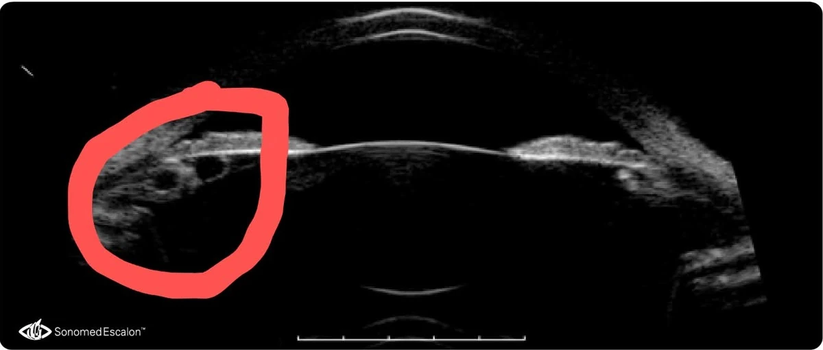
They are iris cysts, small fluid-filled pockets. Many people have them without knowing and they cause no symptoms. They stop being harmless when something has to rest right on top of them.
This patient had them in both eyes. In the left they were small and sat in a way that got in no one’s way. In the right they were larger and sat along the 180-degree meridian, which is exactly where the ends of the lens were going to lie.
A lens resting on a cyst rubs, and from that rubbing come known problems. Pigment released from the iris, chronic inflammation, rotation of the lens with the loss of the astigmatic correction built into it, and the possibility of going back into theatre.
That is why, before implanting, we treated the cysts with laser. A second ultrasound confirmed they were gone, and only then did we go ahead. It is a step that adds a day to the process and can only be taken if someone has looked there first.
What the prediction said about each size
With the ultrasound measurements fed in, the program calculated what would happen with each of the available sizes. This is what came out for the right eye.
| Size | Predicted vault | Predicted angle | Verdict |
|---|---|---|---|
| 12.1 mm | 514 µm | 19.1° | the one chosen |
| 12.6 mm | 986 µm | 12.7° | 77% probability of excessive vault |
| 13.2 mm | excessive vault | 7.1° | ruled out |
| 13.7 mm | excessive vault | 3.9° | ruled out |
The size the external measurement recommended was the 12.6, the second row. On this prediction it would have left the lens almost a millimetre from the crystalline lens and the angle at 12.7 degrees, which is precisely the scenario the three-millimetre threshold exists to avoid.
These numbers have to be read for what they are, estimates from a program and not measurements. The program itself says so and reminds you that it does not replace clinical judgement. And it is worth knowing that what has been published on its accuracy measures the vault, not the angle: in the largest series, of 347 eyes, the predicted vault landed within thirteen microns of the real one on average, and in an independent study the mean error was around 135 microns. But they are estimates that can be compared against what happened next, and that comparison is how this story ends.
The surgery and the result
On 25 August 2026 we implanted both 12.1-millimetre lenses, with reference marks on the cornea to align the astigmatic correction, the haptics placed in the sulcus and the lenses rotated to their axes. Without complications.
The following day, the numbers were these. They come from the first review, at twenty-four hours.
| Right eye | Left eye | |
|---|---|---|
| Uncorrected distance vision | 20/20 | 20/20 |
| Uncorrected near vision | J1 | J1 |
| Vault measured by tomography | ~470 µm | ~470 µm |
| Toric lens alignment | Stable | Stable |
She started seeing 20/400 without glasses and ended up seeing 20/20 with nothing in front of her. That is the jump that explains why she made the trip.
And there are two details worth keeping. The first is that the actual vault was about 470 microns against a predicted 514, with the size that was chosen. Forty-four microns of difference in a measurement that decides whether a lens sits correctly. The second is that at 44, entering the age when near vision starts to cost effort, she kept J1 uncorrected, which is small print without glasses.
What this case does not demonstrate
It is one patient, and one day of follow-up. Both limit what can be concluded, and the second more than the first.
The vault of a phakic lens is judged over time, not in a snapshot, because it changes with pupil dilation and with the years. Pressure and angle in an eye with little room are monitored for years. There is a published case of a highly hyperopic patient whose lenses had to be removed fifteen years after implantation, because of sustained high pressure coming from an angle that had been closing little by little (Cui, Mercieca and Bourauel, BMC Ophthalmology, 2026). It is worth saying that those lenses were of an earlier design, without the central hole the current ones have.
A prediction being right in one eye does not turn it into a measurement. It was right here, with these measurements and in this eye.
Nor does it mean the three-millimetre threshold is superfluous. What this case argues is that an eye below the threshold deserves a look at what is happening inside before being ruled out, not that the threshold stops mattering.
What this changes in clinic
If you have been told you are not a candidate for a lens inside the eye because your anterior chamber is shallow, there are two questions worth asking. Neither disputes the diagnosis; both ask where it comes from.
The first is what they measured the space with. The usual measurement is taken from outside and works well in most eyes. Ultrasound biomicroscopy looks directly at the space where the lens will rest, and sometimes says otherwise. It also shows what lives there, which may be a cyst nobody knew about.
The second is whether that no comes from a figure or from a prediction. They are not the same. A figure below the threshold closes the door without looking; a prediction of how the angle and the vault would end up in your eye opens or closes it with an argument.
The answer may still be no. But it will be a better-founded no.
There is more on how the technique is chosen in refractive surgery with SMILE, and on when a lens inside the eye is the right option in why myopia is more than wearing glasses. Both help explain why the measurements decide more than the prescription does.
Medical note. This article is informative and educational in purpose. It describes an individual clinical case and does not constitute a treatment recommendation, nor does it replace evaluation by an ophthalmologist. The outcomes of phakic lens surgery depend on the characteristics of each eye and are not transferable from one patient to another. The case is published anonymously and with the patient’s consent.
About the author. Dr. Juan F. Batlle Logroño is an ophthalmologist subspecialised in cornea and refractive surgery. He read Medicine at Tulane University School of Medicine, completed his residency in Ophthalmology at Tulane and his fellowship in Cornea and External Diseases at the Bascom Palmer Eye Institute, University of Miami. He practises in Santo Domingo, Dominican Republic.
Sources
- American Academy of Ophthalmology. Refractive Surgery Preferred Practice Pattern. Ophthalmology, 2022, updated 2024. Full text
- U.S. Food and Drug Administration. Summary of Safety and Effectiveness Data, posterior chamber phakic lens. Full text
- U.S. Food and Drug Administration. Approval record for PMA P030016 and its supplements. Record
- Cui L, Mercieca K, Bourauel L. Explantation of implantable collamer lenses due to high intraocular pressure in a highly hyperopic patient. BMC Ophthalmology, 2026. Full text
- Chen X et al. Vault and its influencing factors after implantable collamer lens implantation. Ophthalmology and Therapy, 2021. Abstract
- Gong D et al. Vault after implantable collamer lens implantation. Frontiers in Medicine, 2024. Full text
- Zaldívar R et al. Vault prediction with an artificial intelligence assisted system. Clinical Ophthalmology, 2025. Abstract