A 55-year-old patient came to Centro Láser in April 2026 with a complaint that is more common than it seems: the cataract surgery he had undergone five months earlier had restored his vision, but not the vision he had expected. He saw well at distance (20/25 in one eye, 20/30 in the other), but at near he could barely read at J16. When we evaluated him, we found the reason: he had bilateral residual hyperopia with residual astigmatism and high-order aberrations confirmed by iTrace. The biometric calculation had not been perfect, the EDOF Eyhance lenses that had been implanted are not multifocal lenses (although he had been told they were), and the combined result was vision that worked but did not meet the standard the patient expected.
We proposed a bilateral topography-guided PRK as a bioptic touch-up, an approach that combines the previous intraocular surgery with a secondary corneal laser correction. The day after the procedure he was already seeing at near at J3 without correction (from J16), and distance acuity was maintained at 20/25 and 20/30. This case sums up three lessons worth reviewing seriously when we talk about refractive cataract surgery: the importance of properly managing expectations about the type of lens, the precision of biometric calculation, and the value of the bioptic approach when the refractive result does not reach the goal with intraocular surgery alone.
Why an eye with well-performed cataract surgery may still need glasses
Modern cataract surgery is highly precise, but the intraocular lens (IOL) calculation is a prediction exercise based on biometric measurements of the eye. Variables such as axial length, corneal curvature, anterior chamber depth, and lens thickness are entered into formulas that estimate what lens power should be implanted so the patient ends up with a refraction close to zero.
In most cases, the result falls within an acceptable margin. But not always. A percentage of patients are left with a refractive residual (hyperopia, myopia, or astigmatism) that, though small, limits function without glasses. This is called a post-cataract refractive surprise, and it is a real situation that every surgeon handles with a certain frequency. It is not a surgeon's failure in most cases: it is an intrinsic limitation of biometric calculation in particular eyes.
When the patient also has expectations of complete independence from glasses, the refractive residual becomes a clinical complaint. That is where the bioptic approach comes in: if intraocular surgery has already been performed and left a residual, a corneal laser correction can close the gap between what was achieved and what was sought.
The difference between EDOF lenses and multifocal lenses
This is a point where many expectations get lost in translation. Multifocal lenses and EDOF lenses are two distinct categories of premium intraocular implants, and the confusion between them is one of the most common sources of postoperative dissatisfaction.
Multifocal lenses split the light entering the eye among several discrete focal points: typically one focus for distance, one for intermediate, and one for near. A patient with a well-implanted multifocal lens can read without glasses. In exchange, they may perceive halos or nighttime glare, and distance sharpness is shared among the focal points.
EDOF lenses (Extended Depth of Focus) work differently. Instead of splitting light into several focal points, they produce an elongation of the focus: a continuous range of intermediate vision with less compromise of distance sharpness. In general, they tend to produce fewer halos than high-add diffractive multifocals, although the magnitude varies according to the specific design of the lens. Near vision with EDOF is acceptable for most intermediate tasks (phone, car dashboard, kitchen), but it is usually insufficient for prolonged reading of small print. That is why many patients with EDOF still use reading glasses for certain tasks.
There is also a third important category: enhanced monofocal lenses, an intermediate position between a standard monofocal and a true EDOF. The Eyhance (J&J Vision) belongs to this category. The FDA approved it in 2021 as a conventional monofocal lens, and two recent systematic reviews (BMC Ophthalmology 2023) conclude that it does not meet the ANSI criteria to be classified as a true EDOF. Its design uses a continuous increase in power from edge to center to slightly extend depth of focus, which improves intermediate vision compared to a standard monofocal without generating the halos typical of multifocals. The improvement in near vision exists but is marginal and does not replace the need for reading glasses in most patients.
That distinction (enhanced monofocal, not a true EDOF or multifocal) is exactly what is often not explained clearly to the patient, and it defines whether the postoperative visual result will meet what was expected. In the preoperative consultation, we insist that the patient understand exactly what category of lens will be implanted and what visual range it offers, because managing expectations is half the success of refractive cataract surgery.
The case: 55-year-old man with residual hyperopia post-cataract
The patient had undergone bilateral cataract surgery in November 2025 by another physician, with Eyhance lenses implanted in both eyes. According to his report, he had initially been told they were multifocal. Five months later he came to our practice with visual dissatisfaction, especially at near.
Initial evaluation
The findings were those expected for an eye with a residual post-IOL refraction:
| UCVA distance | Pinhole | Near VA | Subjective refraction | IOP | |
|---|---|---|---|---|---|
| OD | 20/25 | 20/20 | J16 | +0.50 −0.75 × 180 → 20/15 | 10 mmHg |
| OS | 20/30 | 20/25 | J16 | +1.00 −0.75 × 20 → 20/20 | 10 mmHg |
Subjective refraction brought the right eye to 20/15 with +0.50 −0.75 × 180 and the left to 20/20 with +1.00 −0.75 × 20. In other words: with corrective lenses, vision was excellent. Without them, it was sufficient for distance but not for near, and that was exactly what the patient wanted to resolve.
Biomicroscopy confirmed that the intraocular lenses were well positioned in the capsular bag, the anterior segment was healthy, and the fundus showed optic nerves with a 0.1 cup and normal macula, vessels, and periphery. Nothing indicated retinal or posterior segment pathology that would explain the visual complaint.
The diagnosis
The clinical combination pointed to four simultaneous diagnoses:
- Bilateral residual hyperopia
- Residual astigmatism
- High-order aberrations confirmed by iTrace
- Status post EDOF lens implantation (Eyhance)
None of those findings alone justifies clinical concern. Together they explained why a patient with correctly centered IOLs continued to see blurry at near. High-order aberrations are particularly relevant: they are not corrected with conventional lenses, and they limit visual quality even when acuity with glasses appears good.
What the bioptic approach is and why it works
The bioptic is a strategy that combines two procedures in sequence to reach the final refractive goal: an intraocular surgery (phakic lens such as ICL or pseudophakic intraocular lens after cataract) followed by a corneal laser correction (PRK or LASIK) that fine-tunes the refractive residual. The term was originally coined by Zaldivar in 1999 (Journal of Refractive Surgery) to describe the combination of a phakic ICL lens with LASIK in extreme myopias. It was later extended to the pseudophakic post-cataract context, where it is called "pseudophakic bioptic" or "unplanned bioptic" when the corneal correction responds to an unforeseen refractive residual (Moshirfar et al., Expert Rev Ophthalmol 2014).
The logic is the same in both scenarios. The IOL biometric calculation, however precise, never reaches 100% accuracy. Accepting that limitation and planning from the outset for the possibility of a corneal touch-up allows targeting the best combined result, not the best result with a single surgery. In patients who prioritize glasses independence, the bioptic is usually the most reliable route.
In our practice, the subsequent corneal touch-up has another advantage: it also allows correction of the high-order aberrations that remain after an IOL implant, especially with topography-guided technology. That advantage does not exist when only intraocular surgery is performed.
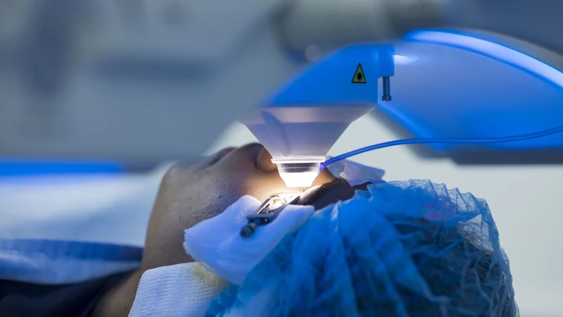
The procedure: topography-guided PRK with Wavelight EX500
PRK (Advanced Surface Ablation) is an excimer laser technique that removes the corneal epithelium and sculpts the underlying stroma to correct the refractive defect. When combined with topographic information (in our case with the Topolyzer), the laser designs a customized ablation profile that addresses in a single treatment both the correction of the refractive error and the regularization of the corneal surface, which reduces the high-order aberrations derived from the patient's corneal geometry.
The specific procedure included:
- Alcohol-assisted manual debridement of the corneal epithelium — removes the epithelial layer to expose the stroma
- Ablation with the Wavelight EX500 excimer laser (Alcon) — the laser sculpts the stroma according to the preoperative plan
- Topography-guided treatment with Topolyzer — the ablation is customized with the patient's corneal map
- Custom calculator — fine refractive adjustment and simultaneous reduction of high-order aberrations
The Wavelight EX500 and Topolyzer are part of the equipment available at Centro Láser, where Dr. Batlle Logroño leads the cornea and refractive surgery program. The combination of excimer ablation with topographic guidance is the current standard for personalized refractive corrections, especially when the eye has documented aberrations.
The surgery was performed under topical anesthesia without complications. The eye was left with a therapeutic contact lens to protect the cornea during epithelial healing.
Results on day one
The day-one check-up showed the functional improvement we were looking for:
| UCVA distance | Near vision | |
|---|---|---|
| OD (dominant) | 20/25 | J3 without correction |
| OS | 20/30 | J3 without correction |
Near vision went from J16 to J3 without correction. The patient was already reporting significant subjective improvement from the first day. Distance acuity remained at pre-PRK values, which is the expected outcome: the goal of the touch-up was to close the hyperopic residual, not to change distance vision.
Biomicroscopy showed what is expected for day one post-PRK: mild conjunctival hyperemia, well-positioned therapeutic contact lenses, a central epithelial defect in the process of healing, clear corneas without opacities, a quiet anterior chamber, IOLs well positioned in the capsular bag, and a fundus without pathological changes.
Complete refractive stabilization after a PRK occurs between 1 and 3 months after the procedure. During that period we perform serial check-ups, remove the therapeutic contact lens once the epithelium has fully healed, and monitor the stability of the refraction to confirm that the result is maintained.
Four lessons from the case
This type of case is seen in the clinic with a certain frequency, and it always leaves the same lessons.
First: managing expectations is half the surgery. The patient had been told his lenses were multifocal when they were actually EDOF. That confusion created a functional expectation that the Eyhance lenses were not designed to meet. Clearly explaining what type of lens will be implanted, what visual range it offers, and what situations may still require glasses is a central part of the preoperative consultation.
Second: biometric calculation precision can be maximized but never reach 100%. Even with advanced formulas and multiple biometric parameters, a small refractive surprise is possible. The goal of preoperative calculation is not to eliminate risk but to minimize it, and to have a plan B when it is not enough.
Third: the bioptic approach is a robust plan B. Combining intraocular surgery with a secondary corneal correction when needed allows targeting the best combined refractive result. Assuming from the outset that the bioptic may be necessary, and discussing it with the patient before the first surgery, is part of mature refractive planning.
Fourth: topography-guided technology optimizes visual quality. It not only corrects the refractive error. It reduces high-order aberrations that limit sharpness even when acuity with glasses is good. For a patient with a suboptimal post-IOL result, that component can be decisive.
When to consult if you are in this situation
If you have already had cataract surgery and the visual result is not what you expected, there are several scenarios where an ophthalmologic evaluation can determine whether a corneal laser touch-up is appropriate:
- Residual blurry vision at distance, near, or both, stabilized at least 3 months after cataract surgery
- Residual astigmatism that was not fully corrected
- Halos, glare, or loss of nighttime sharpness
- Good acuity with glasses but unsatisfactory function without them
- Confusion about what type of intraocular lens was implanted and what to expect from it
The typical evaluation includes full refraction, corneal topography, iTrace aberrometry, biometry, and examination of the state of the IOLs and the retina. Those studies make it possible to decide whether the eye is a candidate for bioptic and what specific corneal procedure (PRK or LASIK) best fits the case.
Frequently asked questions
How long do you have to wait after cataract surgery to perform a PRK?
Generally at least 3 months, to ensure that the refraction and the position of the intraocular lens are stable. In some cases with a very evident and stable refractive residual, it can be considered sooner, but the practical rule is to wait for complete stabilization.
Would PRK damage the implanted intraocular lens?
No. PRK operates on the cornea, a structure completely separate from the intraocular lens, which is inside the eye in the capsular bag. They are two optical tissues in series, and the corneal correction does not affect the position or integrity of the IOL.
Does PRK hurt?
During the procedure, topical anesthesia is applied and no pain is felt. In the first 2 or 3 postoperative days there may be discomfort (foreign body sensation, tearing, sensitivity to light) due to the healing epithelial defect. These symptoms subside once the therapeutic contact lens is placed and are controlled with analgesic and anti-inflammatory drops.
What is the difference between PRK and LASIK in this context?
Both techniques correct the same type of refractive residual. PRK acts directly on the stromal surface after removing the epithelium. LASIK creates a flap and ablates underneath. In post-IOL patients with structurally healthy corneas, both are valid options. The choice depends on corneal thickness, topography, and specific preferences of the case.
How long does the result last?
The refractive result after a well-performed PRK tends to be stable long term. The cornea has limited capacity for regression after the first year, and in most cases the patient maintains corrected vision for many years. However, like any eye, it can continue to change with age, so we recommend annual check-ups.
What this case makes clear
A post-cataract refractive residual is not a dead end. When the biometric calculation leaves a hyperopia or astigmatism that limits function without glasses, the bioptic approach (intraocular surgery followed by corneal laser correction) can close the gap. Topography-guided PRK with current technology not only corrects the refractive error; it also reduces high-order aberrations and optimizes overall visual quality.
The other lesson of the case is preventive: understanding what intraocular lens will be implanted, what visual range it offers, and what expectations are realistic is a central part of well-planned refractive cataract surgery. EDOFs are not multifocals, and each lens category has its specific functional profile. That frank conversation before surgery avoids most subsequent dissatisfactions.
If you had cataract surgery and are not satisfied with the visual result, an evaluation with aberrometry and topography studies can determine whether a refractive touch-up is appropriate for your case.
About the author
Dr. Juan F. Batlle Logroño is an ophthalmologist specializing in cornea and refractive surgery. A graduate of Tulane University School of Medicine and Fellow of the Bascom Palmer Eye Institute, he is Co-Director of CCCRP and of the Dominican Republic Cornea Bank. He practices at Centro Láser, where he leads programs in cornea, refractive surgery, and complex post-laser cataract cases. His practice integrates bioptic surgery and refractive rescue cases, along with cataract surgery in complex post-CAIRS corneas and visual reconstruction in patients with previous penetrating keratoplasty.
Disclaimer
This content is for educational and informational purposes. It does not replace professional ophthalmologic consultation, diagnosis, or treatment. Every case of post-cataract refractive residual requires individualized evaluation, and the appropriate procedure depends on specific anatomical and clinical factors. The results described correspond to a particular patient and may vary according to each person's clinical conditions.
References
- Zaldivar R, Davidorf JM, Oscherow S, Ricur G, Piezzi V. Combined posterior chamber phakic intraocular lens and laser in situ keratomileusis: bioptics for extreme myopia. Journal of Refractive Surgery. 1999;15(3):299-308. PMID: 10367571.
- Moshirfar M, McCaughey MV, Santiago-Caban L. Corrective techniques and future directions for treatment of residual refractive error following cataract surgery. Expert Review of Ophthalmology. 2014;9(6):529-537. PMC4317710.
- Kanclerz P, Toto F, Grzybowski A, Alio JL. Extended Depth-of-Field Intraocular Lenses: An Update. Asia-Pac J Ophthalmol. 2020;9(3):194-202. PMC7299221.
- Megiddo-Barnir E, Alió JL. Positioning of enhanced monofocal intraocular lenses between conventional monofocal and extended depth of focus lenses: a scoping review. BMC Ophthalmology. 2023. PMC10015679.
- TECNIS Eyhance IOL (Model ICB00). J&J Vision Professional. Available at: https://www.jnjvisionpro.com/en-us/products/tecnis-eyhance/
Last updated: April 17, 2026