A patient comes to clinic with a simple question. She wants to be rid of her glasses. Without correction she can make out no more than a hand moving in front of her face, and with the best prescription that can be put in front of her she reaches 20/40 in both eyes, which is half of what a person with normal vision sees.
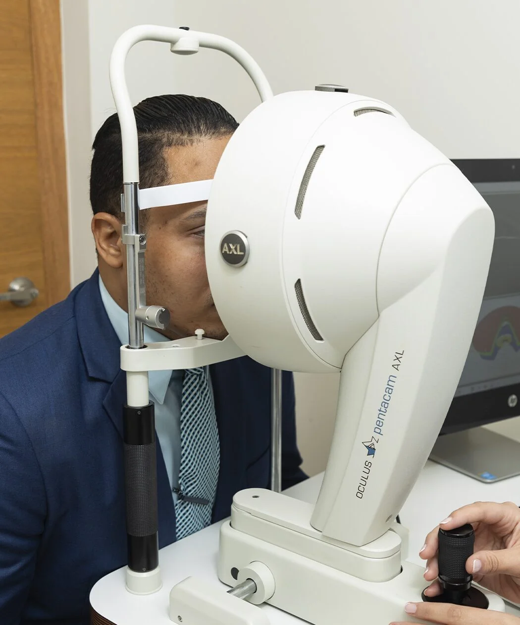
Pentacam tomography explains both findings at once. She has keratoconus in both eyes, with high myopia and irregular astigmatism. She sees poorly, and on top of that the surface that should be focusing the light is deformed, which is a different problem and a harder one to fix.
The answer to her question was that laser was out. The plan that did work had two operations in an order that cannot be reversed. First segments placed inside the thickness of the cornea, which give the surface back a more regular shape, and a little over a year later a lens inside the eye, which corrects the prescription that remained. The order matters because that lens is calculated from the shape of the cornea, and segments implanted later would change that shape and leave the lens prescription worthless.
That sequence is published, and the work closest to this case comes from the cornea unit at Queen Victoria Hospital, in the United Kingdom. They reviewed 14 eyes of 12 patients with keratoconus operated in that same order, the segments first and the toric lens afterwards, and measured what vision and what prescription they were left with at least six months later. All 14 ended with corrected vision of 20/40 or better, when at the start only 3 reached it, and mean refractive astigmatism fell from 4.8 to 1.2 dioptres (McLintock et al., European Journal of Ophthalmology, 2021).
This article describes how that was applied to one case, with what was measured at each step. The two techniques are well known on their own, and what decides the outcome is how they are chained.
Why laser has no place here
Laser corrects the prescription by removing tissue from the cornea. In a healthy eye that is safe, because there is thickness to spare and the structure holds. In keratoconus the situation is the opposite, because the cornea is already thinned and biomechanically weakened, and taking tissue away from it can accelerate the very deformation one is trying to compensate.
That is why the American Academy of Ophthalmology’s practice guideline lists corneal ectasias, keratoconus among them, as a contraindication to refractive surgery (Refractive Surgery Preferred Practice Pattern, 2022, updated 2024). It is one of the contraindications written without qualification, and the reason is the one above, since the procedure removes material from a wall that is already too thin.
There is a distinction worth making, because laser does appear in the management of some weak corneas. What changes is the aim. There it is used to regularise the surface, with very conservative ablations and alongside crosslinking to reinforce the tissue, and the full prescription is deliberately left uncorrected. Two different problems, two different uses of the same machine.
What distinguishes a corneal surgeon is knowing when the answer lies outside the laser. That judgement rests on tomography and corneal thickness, the two measurements that decide whether a cornea will tolerate having tissue removed.
What was measured at the start
These were the starting numbers. They are the reference against which everything that followed has to be read.
| Right eye | Left eye | |
|---|---|---|
| Uncorrected vision | counting fingers | counting fingers |
| Best corrected vision | 20/40 | 20/40 |
| Tomography | significant keratoconus | significant keratoconus |
The line that matters is the second one. With the best possible correction in front of her, this patient saw 20/40, and that was her ceiling. A ceiling of 20/40 in someone of working age means that driving at night, reading a sign at a distance or recognising a face across the street are all tasks that cost effort.
Tomography added the figures used to decide what can be done with a cornea. In the right eye, 426 microns of thickness at its thinnest point and a maximum curvature of 52.5 dioptres; in the left, 424 microns and 54.9 dioptres. Anterior chamber depth, the space where a lens would later have to fit, was around 3.5 millimetres in both eyes.
The reason for that ceiling sits in the word irregular. A lens corrects two things, sphere and cylinder, which describe the simpler distortions of an optical system. When the cornea deforms asymmetrically, as it does in keratoconus, more complex distortions appear that those two numbers do not capture and that spectacles do not compensate. The prescription can be measured perfectly and the image still arrives dirty. Rigid contact lenses do attenuate part of those distortions, because they place a regular surface in front of the cornea, which is why for years they remain the best available correction for many people with keratoconus (Global Consensus on Keratoconus and Ectatic Diseases, Cornea, 2015).
First stage, regularising the cornea
In June 2025, intrastromal corneal ring segments were implanted in the left eye, and in July of the same year in the right. One eye at a time, a few weeks apart, which is the usual practice when it helps to see how the first responds before touching the second.
These are arc-shaped segments placed within the thickness of the cornea, in a tunnel made at the depth calculated for each case. They remove no tissue. What they do is occupy space inside the corneal stroma and shorten the arc of the surface lying in front of them, so that the bulging zone flattens, the tension redistributes and the surface recovers a more regular shape. They exist in two families, made of synthetic material and made of donor corneal tissue, the latter being what we call CAIRS. Mechanically they pursue the same end, and the choice between them depends on the thickness and the condition of each patient’s cornea.
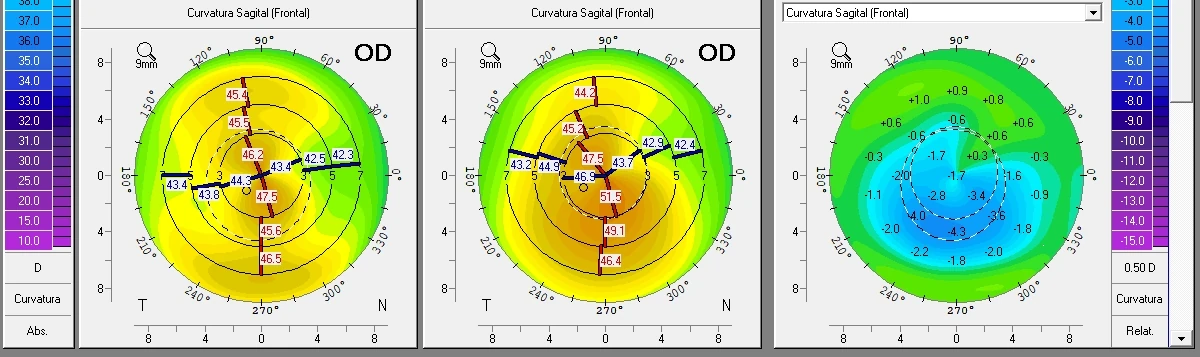
The effect was measured in February 2026, with the tomography in hand. There was a significant reduction in corneal aberrations, regularisation of the irregular astigmatism and central flattening of both corneas.
The flattening was quantified. In the right eye the cornea went from 45.6 and 49.5 dioptres in its two principal meridians to 43.9 and 47.2; in the left, from 45.5 and 50.5 to 43.5 and 47.2. Between two and three dioptres less, in the same direction in both eyes, with the deepest point close to four.
In terms of vision, this is what had changed. These are the numbers from that same February check, with the cornea already settled.
| Right eye | Left eye | |
|---|---|---|
| Uncorrected vision | 20/150 | 20/150 |
| With correction | 20/30 with −6.50 −1.00 × 20° | 20/40 with −5.00 −3.00 × 165° |
There are two readings here and the second is the important one. The first is that uncorrected vision went from counting fingers to 20/150, which in practice means finding your way around a room. The second is that her corrected ceiling rose, from 20/40 to 20/30 in the right eye. That rise comes from the optical quality of a more regular surface, which lets a cleaner image reach the retina to work with.
What remained after this first operation was myopia with astigmatism, high but by then regular. A defect of that kind is correctable, and the question is with what.
Second stage, correcting the remainder without touching the cornea
In August 2026, toric phakic intraocular lenses were implanted in both eyes. Thirteen months had passed since the last corneal procedure, long enough for the corneal shape to settle and the refraction to be stable.
A phakic lens sits inside the eye, in front of the crystalline lens, which is left intact. Hence the name, since phakic means the eye keeps its own natural lens. It is the lens also known as an ICL. Toric means it carries the astigmatic correction built in, oriented along a specific axis, which is why its position inside the eye is planned to the degree.
The advantage in this case is direct. The lens corrects the prescription without removing a micron of cornea, so the structure we had just regularised is left alone. And should it ever be necessary, the lens can be removed or exchanged. That does not return the eye to its starting point in every respect, because a loss of endothelial cells or a cataract already established do not undo themselves when the lens comes out.
There was a surgical detail of the kind one does not anticipate until actually operating. The good optical transparency of the corneal segments allowed adequate intraoperative visualisation. A segment that clouds the periphery of the cornea obstructs the surgeon’s view at exactly the moment when millimetric precision is needed to position the lens, and that was not the case here.
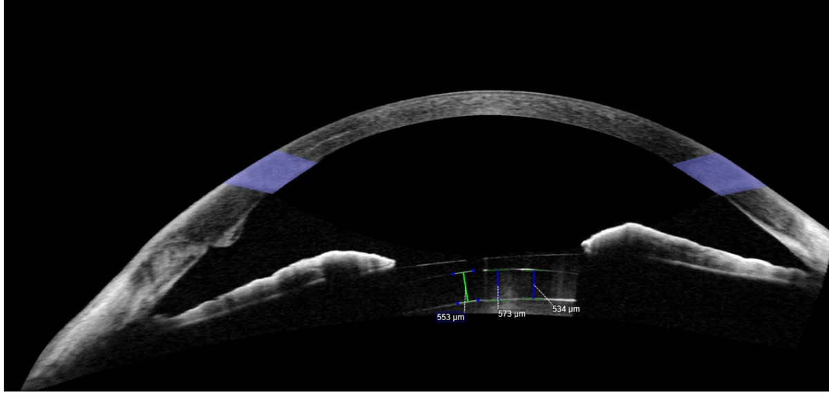
A month later, anterior segment tomography confirmed that the lens had settled where it should. The gap between the lens and the eye’s own crystalline lens, which is watched so that it neither rubs in front nor presses behind, came out between 534 and 573 microns in the right eye and between 615 and 726 in the left.
The result at one month
At one month after the second operation the numbers stood as follows. This is an early check, so what it shows is a snapshot of one moment rather than the final outcome.
| Right eye | Left eye | |
|---|---|---|
| Uncorrected vision | 20/30 | 20/30 |
| Residual refraction | −2.00 −0.75 × 15° | +0.75 −3.50 × 140° |
| Best corrected vision | 20/15 | 20/15 |
The first line has to be read against the starting point. This patient began counting fingers, and a month after the second operation she sees 20/30 with nothing in front of her, better than she saw at the outset with the best correction that could be given to her.
And the third line says something different, which is the finding of the case. Her best corrected vision ended at 20/15, above the 20/20 standard. That figure rearranges the whole case, because the irregular cornea had been hiding a retina capable of 20/15, and the 20/40 of the first consultation was describing the surface more than the potential of the eye.
That rise in the ceiling has two components, and separating them helps to understand the case. One is the corneal regularisation, already visible in February. The other is optical and has to do with where the correction sits. Correcting high myopia with spectacles shrinks the image reaching the retina, and a smaller image is resolved less well. Moving the correction inside the eye removes that effect, and a classic optical model calculated that the gain can reach a line of acuity or more (García et al., Journal of Cataract and Refractive Surgery, 1996). It has to be taken for what it is, a calculation on theoretical myopic eyes of regular optics rather than a measurement in patients with keratoconus, but it matches what we see in clinic every time a high myope compares a contact lens with their glasses.
The myopia that was left on purpose
The right eye was left with two dioptres of residual myopia. That was planned before entering theatre, for a reason that has to do with age rather than with the cornea.
This patient is at the age when presbyopia starts to appear, the point at which arms become too short for reading. A slightly myopic eye focuses up close without help, so leaving that residual in one eye alone buys her years of reading without reaching for glasses. The strategy has a name, monovision, and the figures it is applied with vary. The milder versions work below a dioptre and a half of difference between the two eyes, and beyond that one enters classic monovision, which gives more independence from glasses and demands more adaptation. Most of the literature comes from cataract surgery, and with phakic lenses the published experience is shorter, since the first study to measure it dates from 2017 (Kamiya et al., Scientific Reports).
It does not suit everyone. Some patients do not tolerate the difference between the two eyes, and there are occupations in which that asymmetry bothers more than it helps. It is a conversation held before going into theatre, because afterwards the lens is already inside.
What the literature says about this sequence
The combination of segments first and phakic lens afterwards is published, though in small series and without randomised trials. These are retrospective studies from centres with a high corneal volume, and that is the limitation to read them with.
From the Queen Victoria Hospital series cited at the start, what deserves adding is what they measured uncorrected, which is what matters to someone who wants to stop wearing glasses. Ten of those 14 eyes reached 20/40 or better with no correction at all, starting from zero, and the mean spherical equivalent went from -4.07 to -0.17 dioptres. Its selection criteria are almost as informative as its results. For the segments it required contact lens intolerance, absence of corneal scarring and a thickness of at least 400 microns in the area where they were to be implanted. For the phakic lens it also required a stable refraction, astigmatism already regular on topography, and sufficient endothelial and anterior chamber measurements. Eyes with room to operate, in other words.
Another group followed 16 eyes of 13 patients with moderate to severe keratoconus for two years, in three stages. First the segments, four weeks later the crosslinking, and at least six months after that the toric lens. Uncorrected vision improved from 1.06 to 0.76 logMAR with the first two stages and reached 0.33 logMAR with the lens (Abdelmassih et al., Journal of Refractive Surgery, 2017). At two years uncorrected vision and keratometry were still stable, which is the figure that carries the most weight, because in a progressive disease a first-month result says little.
A recent review of intrastromal segments summarises these studies and adds the appropriate caveat, that only a subset of patients is suitable for this combination, because some eyes still have irregular astigmatism after the segments and in others the corrected vision is not enough to justify the lens (Hayat et al., Clinical Ophthalmology, 2025). It is a narrative review, with no data of its own, and even so that sentence weighs more than any of the figures above.
What does not exist is a study comparing this order against another. In the published series the phakic lens always comes last, with the cornea already stabilised and the refraction settled, so the argument for the order rests on the logic of the calculation and on accumulated practice rather than on a direct comparison.
It depends on the case, and the case is made of measurable things. It depends on corneal thickness, on transparency, on anterior chamber depth, on the endothelial cell count and on the disease being stable.
“The prior corneal regularisation was fundamental in optimising the outcome of the intraocular refractive surgery.”
Dr. Juan F. Batlle Logroño, ophthalmologist subspecialised in cornea and refractive surgery
That sentence sums up the case, and it also orders the conversation with any patient who arrives with the same picture. The first question is whether the cornea can be made regular, and the one about the lens comes after.
What this case does not demonstrate
It is one patient and one month of follow-up after the second operation. In an eye with keratoconus long-term follow-up is not a formality, because the disease can progress and because a lens inside the eye calls for monitoring of the corneal endothelium and the intraocular pressure for years.
The keratoconus was not cured. The shape of the cornea was regularised and the remaining refractive error was corrected. The underlying disease is still there, and the patient will stay under tomographic follow-up.
The result does not generalise. Several favourable conditions coincided here, among them a cornea with enough thickness to house the segments, transparency that did not get in the way during the second operation, and an eye with room for a phakic lens. With any of those missing the conversation is different, and it can end in a transplant or in not operating.
Nor were aberrations measured after the lens. What this case documents is an improvement in visual acuity, not a measured reduction in aberrations in the final state.
What this changes in clinic
If you have been told that you have keratoconus and high myopia, and that you are not a candidate for laser surgery, both statements can be perfectly true. What does not follow from them is that the repertoire has been exhausted.
The useful question is more specific. Whether your cornea can be regularised, with what, and what would be left to correct afterwards. The answer may well be that there is no margin, in which case the path is a different one. But that answer comes from tomography, from corneal thickness and from an endothelial count, not from a prescription. What that workup involves is set out on the page for keratoconus in Santo Domingo.
In my practice the order of these two operations is not negotiable, and the reason is arithmetic before it is surgical. On an irregular cornea, the number one measures in order to calculate a lens does not describe the eye well. Regularising first changes that number, and it is from the new number that the lens is calculated. Doing it the other way round leaves the patient with a well-implanted lens and vision below what was within reach.
There is more on the myopic eye and its lifelong risks in why myopia is more than wearing glasses, and on donor tissue segments in the article on CAIRS. Both help explain why high myopia is monitored for life even after it has been corrected.
Medical note. This article is informative and educational in purpose. It describes an individual clinical case and does not constitute a treatment recommendation, nor does it replace evaluation by an ophthalmologist. The outcomes of keratoconus surgery and of phakic lenses depend on the characteristics of each eye and are not transferable from one patient to another. The case is published anonymously and with the patient’s consent.
About the author. Dr. Juan F. Batlle Logroño is an ophthalmologist subspecialised in cornea and refractive surgery. He read Medicine at Tulane University School of Medicine, completed his residency in Ophthalmology at Tulane and his fellowship in Cornea and External Diseases at the Bascom Palmer Eye Institute, University of Miami. He practises in Santo Domingo, Dominican Republic.
Sources
- American Academy of Ophthalmology. Refractive Surgery Preferred Practice Pattern. Ophthalmology, 2022, updated 2024. Full text
- Gomes JAP, Tan D, Rapuano CJ et al. Global Consensus on Keratoconus and Ectatic Diseases. Cornea, 2015. Full text
- McLintock CA, McKelvie J, Miraflores Gomez S et al. Outcomes of sequential intracorneal ring segment and phakic intraocular lens insertion for visual rehabilitation in keratoconus. European Journal of Ophthalmology, 2021. Abstract
- Abdelmassih Y, El-Khoury S, Chelala E et al. Toric ICL implantation after sequential intracorneal ring segments implantation and corneal cross-linking in keratoconus: 2-year follow-up. Journal of Refractive Surgery, 2017. Abstract
- García M, González C, Pascual I, Fimia A. Magnification and visual acuity in highly myopic phakic eyes corrected with an anterior chamber intraocular lens versus by other methods. Journal of Cataract and Refractive Surgery, 1996. Abstract
- Hayat UK, Shiwani HA, Memon D, Walkden A. Intracorneal ring segments in keratoconus: a narrative literature review. Clinical Ophthalmology, 2025. Full text
- Kamiya K, Takahashi M, Takahashi N, Shoji N, Shimizu K. Monovision by implantation of posterior chamber phakic intraocular lens with a central hole for early presbyopia. Scientific Reports, 2017. Full text